Case studies
Mr Venki Sundaram’s cataract surgery is available in clinics in Harpenden, Bushey and Luton. Contact us today to find out more about the process of cataract surgery or to book an initial consultation.
-
Vic Brown had been having trouble with his vision for a long time and had needed to wear glasses for both short and long distances for over 30 years.
As you can imagine, Vic’s vision slowly deteriorated over this time. This deterioration meant that Vic wasn’t able to enjoy doing the things he loved most during his retirement, so he decided to get his eyes checked to find out what the issue was.
He was quickly made aware that both of his eyes had begun to develop cataracts.
“I had lost a great deal of vision in one eye due to a cataract and the second eye was also developing a cataract. I was finding it increasingly more difficult to see”, explained Vic.
After hearing this diagnosis, Vic got in touch with his insurers who recommended Mr. Venki Sundaram without hesitation. After meeting each other for the first time in person, Vic was very impressed with Mr Sundaram and knew he was is in safe hands from the beginning.
“When I first met him, I was instantly impressed with his manner and professionalism which gave me great confidence.”
The procedure
Vic underwent cataract surgery to remove cataracts in both of his eyes in April and May 2021. Considering that the procedures took place over lockdown, Vic was impressed with the planning of the surgery to make sure the correct protocols were followed.
“This whole procedure was during the latest Covid ‘lockdown’, and everything was very well managed and appointments including surgery went smoothly as planned.”
Before going into the operation, Vic admits that he was a little concerned, but was never scared thanks to Mr Sundaram’s professionalism.
“Not once was I scared as Venki and his team were so professional and gave me every confidence that they knew exactly what they were doing at all times.”
When asked about how the operation went, Vic was surprised at how smooth it was. He was also very impressed at how quick it was.
“Both operations went very smoothly and were quicker and not as intrusive as I had imagined.”
Vic explained how it didn’t take long for him to recover from the operation. In fact, he was able to see the improvement in his vision straight away.
“Immediately after the operation was over, I was able to see without the aid of glasses which was a major improvement within minutes. I followed the program of applying eye drops for a period of four weeks on each eye and everything went to plan.”
The results
Now retired, Vic Brown wanted to enjoy his retirement by getting the most out of what he loves doing most in life. However, he wasn’t able to do that due to the troubles he had with his vision. Thanks to his cataract surgery being a huge success, the results of the procedure have given him much more freedom in life.
“Not having to wear glasses has given me so much more freedom. The brightness and clarity of my vision has been quite exhilarating and life enhancing.”
Unsurprisingly, when asked whether he would recommend Mr. Venki Sundaram as an eye surgeon, Vic’s answer was resounding: “absolutely.”
As a result of Mr Sundaram’s excellent work, Vic expressed how it is now a “pleasure to do virtually all everyday things” without having to wear glasses. Vic is now able to play tennis and golf as well as driving his car without them – and he’s looking forward to enjoying his retirement with improved vision!
-
Having worn contact lenses and glasses since an incredibly young age, Jacqueline Willis has struggled with her vision for most of her life, and in April 2020, Jacqueline underwent surgery to repair a detached retina.
Unfortunately, a cataract in her left eye developed quite quickly after this surgery and her sight began to deteriorate. This affected her ability to read and use the computer and iPad, causing problems in her job as an Early Years Keyworker.
“Part of my work requires me to record observations on the children using an iPad. This was hard as I could barely see what I was typing. When reading a story to a class of 40 children, I found it difficult to hold the book up so the children could see, as I could not see the print.”
Jacqueline decided something needed to be done in order to help her at work. She got in touch with her insurance company and Mr Sundaram’s name was recommended. She explains, “when I got off the telephone, I looked up Mr Sundaram’s profile and was satisfied with my decision especially as he had trained at Moorfields.”
After agreeing to be treated by Mr. Venki Sundaram, Jacqueline was very impressed with his service.
“During my first consultation, Mr Sundaram was professional but also friendly and reassuring making me feel comfortable and able to ask questions. On my second visit I was impressed that Mr Sundaram remembered where I worked, showing that he has an interest in his patients.”
The procedure
Jacqueline had cataract and replacement lens surgery. After having a bad experience the last time she had surgery, Jacqueline understandably had her doubts.
Prior to the operation, she was a bit apprehensive about not having a sedative, but Mr Sundaram reassured her concerns.
“Before the operation, I was a little unsure about not having a sedative. Mr Sundaram reassured me that it was not necessary explaining what anaesthesia would be used.”
She also added that “on the day of the operation Mr Sundaram came in before the procedure again asking if I had any questions”, ensuring everything was in place to help her feel at ease. Jacqueline went on to explain how the procedure went “very smoothly.”
When discussing how long it took to recover from the surgery, she explained how she “was surprised at how quick the procedure was.” Jacqueline also added that she was able to recover from the surgery “within 24 hours”, adding that “it was a little painful during the first night” but was “all fine” the next day.
The results
The results of the procedure have helped Jacqueline Willis enjoy watching TV, swimming, reading and driving once again as well as allowing her to appreciate the little things in life.
“It’s the day-to-day things that other people take for granted like being able to see the clock/watch when you wake up, seeing signs in shops, the number on the petrol pump when you are paying, putting make up on, doing your hair, I could go on and on…”
When asked if she would recommend Mr. Venki Sundaram as an eye surgeon, Jacqueline replied with no hesitation; “Yes, definitely.”
Thanks to his fine work, Jacqueline no longer needs to run around trying to find her reading glasses to see the computer screen at work anymore.
“My vision is so much better. Having worn contact lenses and glasses since a very young age I had become used to not seeing as well; the vision was ok but now everything is so much sharper.”
-
Haydn Thomas’s had been suffering with his eyesight for some time. He first had to wear glasses ten years ago for close up work, such as reading or using the computer. Since 2018, he has needed to introduce glasses for day-to-day activities such as driving, watching TV and taking part in physical activities, such as skiing.
Eventually, Haydn’s eyesight had begun to deteriorate to the point where it was not safe to drive, difficult to work and general activities had become potentially dangerous.
In February 2021, Haydn Thomas was diagnosed with cataracts.
As a result of this diagnosis, Haydn was advised to refrain from driving with immediate effect, and as an active 54-year-old in the process of setting up a new business, Haydn quickly realised that he would need medical intervention in order to maintain and improve his way of life.
Haydn had a general idea of the procedure that would be needed, and had undertaken research into the best eye surgeon for him. Quickly settling on Mr. Venki Sundaram, Haydn never looked back.
“From the first meeting to the last check-up, I found Mr. Venki Sundaram to be extremely efficient. He explained each step and welcomed and answered my concerns and questions.”
The procedure
Haydn had cataract surgery, with two procedures separated by five days, to replace the lenses in both eyes with multifocal lenses. Both procedures were day procedures and took no longer than three hours from arrival to departure.
“During the two days I had the procedures I found the staff at the clinic to be friendly, polite and considerate. The administration of the practice was efficient, notifying me early of appointment dates and arranging Covid tests,” said Haydn.
Within a day of each operation, it was possible for Haydn to remove the clear Perspex coverings and he felt very little discomfort. The remaining discomfort, which he described as “negligible”, took two to three days to subside so it was not noticeable. After a week, his eyes had really settled back. There was a course of four weeks of drops to follow up (From 12 drops to four a day).
“What I do want to impress”, commented Haydn, “is that as soon as I came out of the procedure, I could notice a difference. Whilst I had a clear Perspex cover over the eye, I could look through the holes and it was possible to see the change.”
When asked how Haydn found the whole experience, his response was;
“The procedure is quick (mine were no more than 10 minutes each) and it is not without a little discomfort, as with any surgery. However, the benefits it brings far outweigh anything I can imagine… it is truly life changing. If your eyesight can be corrected using this procedure then I would recommend doing it, even if the only benefit is not to have to wear glasses again. I am in constant awe that Dr. Venki Sundaram conducts this procedure many times a week and makes such a difference to so many people’s lives.”
The results
Like many people, Haydn Thomas found that the impact of the global pandemic had led to him re-evaluating his life. The results of the procedure have now enabled him to make significant decisions on his professional future. He can also enjoy cycling, watching TV, cooking, reading and driving again!
“I have no hesitation in recommending Mr. Venki Sundaram to anybody considering this procedure. The outcome of my procedures has been excellent and I was looked after at each stage of the process. I can now see clearly and do not need glasses. My vision is effectively back to where it was when I was a teenager. I can see long and short distance with impressive detail.
“Everything that I used to avoid I can now do without worry. I can confidently undertake tasks which I had avoided and can also drive again. It is almost two months since the operation and I am still noticing improvements and I can honestly say I wake up every morning, astonished by the detail I can see. I am very thankful for this procedure and I am amazed by the improvement that has been made.”
-
As a director in a broadcast company, Kevin’s career is largely orientated around work on PCs and his eyesight is crucial.
Kevin had a cataract in his left eye, meaning that his left eye was out of focus for longer distance sight. This was beginning to make driving very difficult for him, as well as having an impact on his ability to carry out general day-to-day tasks.
After seeking advice from his medical aid, BUPA, he was given a list of recommendations of their preferred surgeons. Along with his wife, he looked at their profiles online and they opted for Mr Venki Sundaram to carry out the surgery. Kevin commented, “when looking at his website, he had a lot of experience.”
We caught up with Kevin to hear about his experiences with surgeon, Mr Sundaram, and his thoughts post-operation.
On the day
In August 2019, aged 60, Kevin underwent cataract surgery in his left eye. Like with all surgeries, Kevin was a little worried. However, he described Venki as “very pleasant and a really nice person”, with the suggestion of a semi-anaesthetic by Venki to help him not feel so stressed.
Following a smooth operation, “within 24 hours I could not believe the improvement in my sight”, Kevin said.
Kevin explained how it took him just a few days to recover from the surgery, with his eye feeling a little scratchy for just four days post-surgery. But after a speedy recovery, overall, he has seen “massive improvement” to his vision since the surgery.
The results
Kevin’s new and improved vision has helped to improve his quality of life.
He went on to explain the personal benefits this has had: “I now have proper long-distance vision in both my eyes that is great for day-to day-things like driving, walking and cycling.” As a keen cyclist, Kevin has found that surgery has vastly improved his sight for when he’s out cycling.
When reflecting on the surgery, Kevin said “I was very happy with my surgery. I would 100% recommend Venki Sundaram as an eye surgeon.”
Finally, Kevin went on to say how much of an improvement the successful surgery has had. “It’s really been an ‘eye opener’ to have the surgery and I really didn’t think my sight would be that great post-op”, he says.
-
Housewife and former dental nurse Anita Cullen had had a cataract for many years. At first, this only affected her vision in small ways, but as time went by it became progressively worse. At the age of 61, it was getting to the stage where her eyesight was severely affected, leaving her almost blind in her right eye. As her condition started to impact on daily life, it was time to think about eye surgery.
Anita searched online and came across Mr Venki Sundaram on the Spire website. “I chose Mr Sundaram because I was impressed by his resume,” she explains. Specialising in cataract surgery, the consultant ophthalmic surgeon had the qualifications and expertise she was looking for to treat her right eye.
Another thing she valued was the speed at which she could be seen. “I was able to see him quickly, which was important because the cataract deteriorated around Christmas time and was giving a lot of discomfort,” she remembers. Anita’s cataract surgery was scheduled for January. Six months later, we caught up with Anita to find out how this operation has changed her life.
On the day
In January 2019, Anita underwent cataract surgery with Mr Venki Sundaram. Cataract surgery is a common procedure with a high success rate, but the prospect of any operation can be nerve-wracking. Luckily, Anita’s apprehensions were put to rest by her surgeon and the team. “I was nervous, Mr Sundaram was calm and reassuring,” she says.
Because cataract surgery is a micro-incisional procedure, there’s usually no need to go under general anaesthetic. All that’s normally needed is an anaesthetic eye drop to numb the feeling in the eye, which Anita had.
After a painless procedure that took just 10 minutes, it was all over. “The operation went smoothly, and my experience was excellent,” reports Anita. She was allowed to go home on the same day. After this, everything progressed normally, as Anita’s eye slowly healed. “My recovery took approximately 2 weeks,” she remembers.
The results
Six months on, and Anita’s eyesight is now clear in both eyes, allowing her to carry on with daily activities as normal.
“Following the operation, my vision is perfect in the right eye,” reports Anita. “I can now see everything much more clearly, which has improved my life greatly.”
Anita was careful to follow all the aftercare advice offered by Mr Sundaram, which included using eye drops as instructed, using an eye shield at night time for one week , and going without makeup for 4 weeks following the operation. This allowed her right eye to recover fully.
It took years for Anita to go for surgery, but her vision was transformed in just 10 minutes. At the end of her patient journey, would Anita recommend Mr Sundaram to others?
“Yes, I would definitely recommend Mr Sundaram, he was very helpful and the result has been excellent,” she says.
-
At 56 years of age, Steven’s cataracts were beginning to severely affect his life and he was told by his optician that they would potentially cause him to go blind within 12 weeks. After receiving this frightening news, Steven immediately started looking into surgery. As an Architectural Technologist and Senior Design Manager, Steven’s career is heavily dependent on his eyesight, especially when looking at document plans as well as attending site inspections for potential projects, so there was a lot at stake.
To avoid the long waiting list that often comes with the NHS, Steven opted to go private, deciding on The Spire and surgeon, Mr Venki Sundaram, down to them both having an excellent reputation and being easily accessible to him from home.
We caught up with Steven to hear more about his surgery journey as well as his thoughts and feelings on the process and surgeon, Mr Sundaram.
On the day
In the lead up to the surgery and on the day, Steven was naturally a little nervous. However, these nerves were soon alleviated. “I don’t like touching my eyes in any way and to date didn’t even like eye drops. The team was very calming and the whole team explained the stages clear slowly and made sure I understood what was happening and why.”
Following the successful surgery that only lasted 20 minutes in total, Steven was walking out of the hospital within 30 minutes and after a couple of days, he was “100% fine”.
Although his eyesight was fully functioning within a few hours, Steven decided to take a few days’ rest in order to ensure a smooth and relaxing recovery.
The results
Prior to his cataract surgery, Steven’s eyesight had a prescription of +4.5 which has now improved significantly. Steven previously required the constant assistance of a +1.5 reading lens and a blue screen filter. However, his improvement means that this is no longer the case. “My vision is 95 % now, improved beyond scale” explains Steven. “The improvement is beyond words, removing the need for glasses all the time, only for close computer work.”
As well as a massive improvement in his ability to work, a personal benefit has been being able to drive again as he found that the car headlights were no longer “totally blinding” and has enabled the lesser use of yellow lens glasses.
Steven has now regained fully fledged confidence and ability. He went on to explain how the successful surgery has had a positive impact, saying that “this treatment has given me back years of my life, returning my confidence and ability to conduct myself normally when outside the home environment.”
Reflecting on his life-changing surgery and his treatment by Venki Sundaram, Steven said that “Mr Sundaram’s friendly, approachable, relaxed and non-hurried nature, leaves me with no reason not to recommend him.”
-
This is a loss vision that occurs either immediately or within a few minutes. This acute loss of vision is usually due to a problem with the blood supply to the retina and occurs in conditions such as Central Retinal Artery Occlusion, Central Retinal Vein Occlusion, Wet AMD. Sudden loss of vision can also occur following a blockage of blood supply to the optic nerve.
-
This is a loss of vision that typically occurs over several weeks to months. The most common cause is a cataract, but can also occur in Dry AMD.
-
A sudden episode of brief flashes of lights and floaters is usually caused by a posterior vitreous detachment (PVD). This is a natural, age-related process in which the vitreous jelly separates from the retina. Occasionally this process can lead to a retinal tear or retinal detachment occurring.
-
This is a common complaint and can be caused by eye infections (e.g. conjunctivitis), any injury or irritant to the eye, inflammation of the eye or sometimes burst blood vessels on the surface of the eye. In contact lens wearers, a serious cause is a corneal ulcer that needs to be treated promptly.
-
This most commonly occurs when there is a problem with the blood supply to the eye muscles that control the eye movements, and are called nerve palsies. Thyroid problems or inflammation around the eyes can also cause double vision. Occasionally, patients complain of double vision which is only present when 1 eye is open and this can be caused by a specific type of cataract.
-
This is a common complaint and can be due to allergies, dry eyes (which stimulate an overproduction of tears), irritants on the eye, abnormalities of the eyelids and problems with the drainage of tears from the eye.
-
This is usually caused by an irritation or injury to the cornea (which has many nerve endings) through foreign bodies, trauma and dry eyes. Inflammation within or around the eye in conditions such as iritis and scleritis can also cause painful and red eyes.
General Eye Complaints
The eye can be affected by many conditions which can cause a range of symptoms and complaints. Assessment by an Ophthalmologist is generally advised as there may be a number of conditions that can lead to similar symptoms, which may only be diagnosed and managed with specialist examination, assessment and treatment.
Investigations
There are several investigations important that are used in Ophthalmology to help diagnose and monitor eye conditions.
Retinal Photography
This involves using a special camera that allows accurate colour documentation of how the retina appears, which can be useful in documenting conditions and monitor any progress over time.

An example of a colour retinal photograph in a patient with advanced AMD
Optical Coherence Tomography (OCT)
OCT has transformed the understanding and treatment of retinal conditions in recent years. It is a non-invasive technique that captures specialised image of the retina, allowing individual retinal layers to be viewed. It is particularly useful in detecting fluid or bleeding that may occur in the retina in conditions such as Wet AMD, Diabetes and Retinal Vein Occlusion.
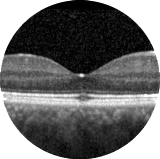
An example of an OCT scan showing the different layers of the retina.
Autofluorescence (AF) Imaging
This is a non-invasive imaging technique that captures naturally emitted light wavelengths from the retina. It allows the detection of areas of retina that are damaged, and is useful in monitoring progression of retinal diseases.

An example of AF imaging in a patient with a condition affecting both retina
Fluorescein Angiography (FFA) and Indocyanine Green (ICG) Angiography
These are common diagnostics tests that involve the injection of a fluorescein (yellow) dye and/or indocyanine green dye into your bloodstream, through a vein in your arm. A series of photographs of the back of the eye are then taken, which can provide information on the blood supply affecting the retina, or the choroid – which is the blood supply beneath the retina. This is particularly useful in helping to diagnose leaking blood vessels in Wet AMD, and the extent of reduced blood supply in conditions such as diabetic retinopathy.
What are the risks of FFA or ICG Angiography?
1 in 10 patients may feel slightly nauseous, short of breath or develop a transient rash, but this rarely lasts for more than a few seconds.
The fluorescein dye can give your skin a yellow tinge and make the colour of your urine yellow, but this usually settles after a day or two.
There is an extremely rare (less than 1 in 200,000) risk of a severe allergic reaction to these dyes.

An example of an FFA image, show details of the retinal blood supply.
Visual Field Testing
This assesses the extent of your peripheral field of vision, which can be affected in conditions such as glaucoma. The test involves responding to small flashes of light which are targeted at various part of your peripheral vision and a map of your visual field is established.
The human eye is an incredible organ that is responsible for the conversion of light signals into meaningful images that can be interpreted as vision by the brain. There are several structures within the eye that have a vital role in this process (see Image).
About the Eye
The human eye is an incredible organ that is responsible for the conversion of light signals into meaningful images that can be interpreted as vision by the brain.
There are several structures within the eye that have a vital role in this process (see Image 1):
Cornea – this is the clear window at the front of the eye, which allows light to enter.
Iris – this is the coloured part of the eye with the hole in the middle known as the pupil.
Lens – this is a clear, convex structure within the eye that helps to focus images onto the retina.
Vitreous – this is the clear, jelly like substance that forms the majority of space within the eye.
Retina – this is the light sensitive tissue at the back of the eye that captures light signals and coverts them into electrical impulses that are sent to the brain. It is the most active tissue in the human body, responsible for processing millions and millions of pieces of visual information over a lifetime.
Macula – this is the central part of the retina and is responsible for central vision.
Fovea – this is the very centre of the macula area and is responsible for providing fine vision.
Choroid – this is a rich network of blood vessels that provide essential oxygen and nutrients to the retina.
Optic nerve – this is the main nerve that leaves the back of the eye and conveys electrical signals from the retina to the brain.
Sclera – this is the tough, white outer coating of the eye that helps to maintain its shape.
