Retinal Therapies

Diabetic Eye Disease
Diabetes is a condition in which there are increased levels of sugar (glucose) in the blood. Consistently high blood sugar can lead to damage of retinal blood vessels, which can lead to diabetic eye disease in the form of diabetic retinopathy (DR) and diabetic macular oedema (DMO).
-
Diabetic retinopathy (DR) occurs as a result of blood vessel damage in the peripheral retina. Left untreated, fragile new blood vessels can grow in the retina. These have a tendency to bleed further, eventually causing loss of vision and scar tissue. For this reason, is it crucial to seek diabetic retinopathy treatment as soon as you are aware that you are affected by a more severe case.
-
Initially, you may experience small, retinal haemorrhages. However, this can progress to more widespread retinal bleeding with larger areas of the retina becoming starved of oxygen.
Retinal Vein Occlusion
Retinal Vein Occlusion is when a blockage occurs in the eye, causing sudden but painless loss in vision. The retina is the thin lining at the back of the eye that allows us to see. When this becomes disturbed due to a blockage in one of the veins, it can disrupt the cells we use to detect light and begin causing serious problems within the eye.
There are two types of this condition, Central Retinal Vein Occlusion (CRVO) and Branch Retinal Vein Occlusion (BRVO).
-
Central Retinal Vein Occlusion is when a blockage occurs in the main central vein in the eye. When affecting this part of the eye, loss of vision tends to be more severe.
-
Diabetes
Hypertension
Raised cholesterol
Smoking
Raised eye pressure
Cardiovascular Atherosclerotic Disease
Typically, CRVO occurs in patients over 50 year of age. If the condition occurs in younger patients, this is more likely to be as a result of an underlying blood disorder.
-
Symptoms include a painless but sudden reduction in vision.
Complications of CRVO include;
Growth of abnormal blood vessels – these fragile blood vessels can grow in the retina or iris, causing bleeding, further vision loss or glaucoma.
Macular oedema – a thickening and swelling of the macula can occur as a result of damage to the retinal vessels causing leaking and blurring or distortion of vision.
-
Branch Retinal Vein Occlusion occurs when there is pressure on one of the four retinal veins, from an overlying artery. There are similar underlying factors associated as there are with CRVO, however, it is actually three times more common.
-
Loss of vision – patients may experience a loss in part of their peripheral vision where the BRVO is located.
Abnormal blood vessels – these can develop in the retina which may bleed if left untreated.
Macular oedema can develop, significantly affecting vision.
-
Unfortunately, there is currently no treatment available to reverse the blockage of the vein. However, symptoms can be managed and underlying issues such as diabetes, blood pressure or cholesterol issues and raised eye pressure can be better controlled to prevent further complications.
The following methods are used to manage both Central and Branch Retinal Vein Occlusion and prevent the condition occurring in the other eye:
Careful monitoring
Regular monitoring to assess growth of abnormal blood vessels is implemented, in order to avoid further vision impairment. If this does occur, laser treatment (PRP) can be given.
AntiVEGF therapy
AntiVEGF therapies such as Avastin, Eylea and Lucentis have been proven to treat macular oedema caused by RVO. Carried out with a course of injections, patients have seen a reduction in macular oedema and an improvement in vision.
Steroid therapy
Ozurdex is a type of steroid therapy involving a steroid implant being injected into the eye, proved to treat macular oedema due to RVO.
Macular laser
Small, light laser spots are applied to the area of macular swelling and can be effective in the reduction of the oedema and restoration of vision.

Retinal haemorrhages and damage
caused by a CRVO

Macular oedema due to a CRVO
Myopic Degeneration
Myopic degeneration is the damage caused to the retina as a result of abnormal blood vessels and stretching of the eye due to high myopia (also known as short sightedness).
Myopia is a condition where the eyeball grows to a greater length than usual, causing individuals to see close objects clearly but distant objects appear blurry.
The stretching of the eye and thinning of the retina that occurs can lead to bleeding and leaking of fluid into the retina, negatively affecting vision.
-
Main symptoms include;
Reduced vision
Distortion of images – blurring/spotting
People with high myopia are more susceptible to other eye conditions, such as retinal detachment and glaucoma, so should have regular check-ups and keep an eye out for any sudden changes in vision.
-
Myopic degeneration is diagnosed by ophthalmic examination including retinal imaging.
-
This type of diabetic eye disease tends to occur at a later stage, with approximately 10% of diabetic retinopathy patients developing diabetic macular oedema. Caused by damage to the fine retinal blood vessels in the macular area of the eye, it can lead to leakage of proteins and fluid into the retina, as well as a reduction in vision. Left untreated, irreversible vision loss can occur due to permanent damage to the retinal photoreceptor cells.
In fact, diabetic macular oedema is one of the leading causes of visual impairment in the working age.
-
AntiVEGF therapy can be used to target the abnormal blood vessels and any bleeding or leaking that has occurred in the eye as a result of myopic degeneration. It has proven to be extremely effective in the improvement and prevention of vision loss.
This treatment involves an injection of AntiVEGF (such as Avastin, Eylea or Lucentis) into the vitreous jelly in the eye.
The procedure as a whole is only around 5 minutes long with the patient being able to return to normal activities immediately.
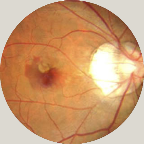
An illustration of retinal bleeding
at the macula in a patient with high myopia
AntiVEGF Therapy
AntiVEGF therapies have been used to treat retinal conditions for the last few years, revolutionising the vision and lives of many patients.
In several conditions such as; Wet AMD, Diabetic Macular Oedema, Retinal Vein Occlusion and Myopic Degeneration, there is bleeding or leaking from abnormal or damaged retinal blood vessels.
-
AntiVEGF therapies work by targeting these abnormal blood vessels, reducing or stopping the bleeding and leakage that can occur. This typically results in prevention of further loss of vision, and can also lead to improved vision.
-
There are currently 3 types of AntiVEGF therapy:
Eylea and Lucentis – this is licensed to be used in Wet AMD, Diabetic Macular Oedema, Central and Branch Retinal Vein Occlusion and Myopic CNV Degeneration.
Avastin – this is not licensed for use in the eye, but was the first type of AntiVEGF therapy to be delivered, and is the most widely used AntiVEGF agent in the USA.
The type of AntiVEGF therapy used depends on factors of the individual and their existing treatment plan. Most will require a course of 3 monthly injections with further potentially given, depending on the response to initial therapy.
-
All AntiVEGF treatments are given as injections into the vitreous jelly of the eye and done as an outpatient procedure.
The process:
The eye is numbed with anaesthetic eye drops.
The skin around the eye is cleaned and a sterile drape is placed over it, then a small clip, known as a speculum, is inserted to keep the eyelids open.
0.05ml of AntiVEGF is injected into the eye using a small needle. (This should not be painful due to the anaesthetic administered prior)
Antibiotic drops are applied, with the patient continuing this for the following days after.
The whole procedure takes around 5 minutes with the injection lasting less than 20 seconds.
Patients can generally return to normal activities straight after the procedure.
-
With an excellent safety profile, there is very minimal risk involved in the procedure.
Minor side effects that may occur:
Red eye – this is usually due to bleeding of tiny blood vessels on the surface of the eye and resolves by itself within a few days.
Blobs in the vision or flashing lights – this is caused by a slight disturbance of the vitreous jelly as the injection is given and normally resolves within a few hours.
Sore eye – the eye may be slightly achy for a day or two following the injection.
With anything, there are potential risks however these more serious complications occur in less than 1 in 1000 patients.
This could include:
Serious infection in the eye
Detached retina
Raised eye pressure affecting vision
Bleeding in the eye
Inflammation in the eye
Cataract
-
The AntiVEGF injection is not advised when:
There is a history of allergy to a particular AntiVEGF therapy.
A patient is trying to become pregnant, is currently pregnant or is breastfeeding.
There is a history of heart attack or stroke in the last 3 months.

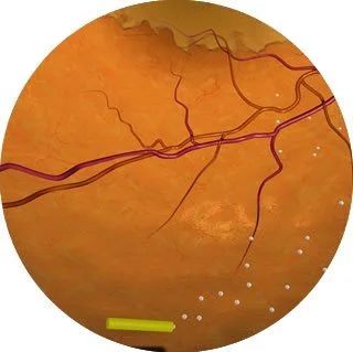
An illustration of an AntiVEGF injection being given into the eye
Retinal Laser
Laser is a common treatment method within the eye for visual impairment and complications caused by conditions such as diabetic retinopathy or retinal vein occlusion. It often works by targeting and reducing the growth of abnormal blood vessels or fluid leakage. Different types of retinal laser are recommended for different conditions and symptoms, these are; pan-retinal photocoagulation (PRP) and macular laser.
-
PRP is a type of laser treatment, used specifically when patients have developed abnormal blood vessels in the retina. This often occurs as a result of severe diabetic retinopathy or following retinal vein occlusion causing an impairment in the blood supply to the retina. To avoid permanent damage, PRP is recommended.
-
PRP works by a laser targetting these new blood vessels and encouraging them to shrink and scar in order to prevent more from growing, and helps avoid bleeding into the vitreous jelly of the eye. Without this treatment, there is a risk of loss of vision if the condition develops.
-
Macular laser is a type of treatment used to treat leakage of fluid from the macula region in the retina as opposed to growth of blood vessels. The macula is part of the retina that is responsible for detailed vision therefore any fluid that leaks into this area can lead to a loss of central vision. This can occur as a result of conditions such as Diabetic Macular Oedema and Branch Retinal Vein Occlusion.
-
Small lasers help dry up and reduce leakage of fluid to this area in the eye. This is strongly advised and if not treated, damage can be caused to the retina, permanently affecting your central vision.
Macular laser will prevent vision from getting worse with some patients even experiencing improved vision following this treatment.
-
The procedure on the day is very similar to that of pan-retinal photocoagulation. The differences are that a slightly different contact lens is used, in order to gain a highly magnified view of the macula when carrying out the treatment. Also, a fewer number of laser spots are needed as well as a lower laser intensity.
Please be aware that further laser treatment may be necessary in the future, depending on the outcome of your 3-month follow up assessment.
-
Pan-retinal photocoagulation laser is carried out as an outpatient procedure at one of our Hertfordshire clinics by the trusted surgeon, Venki Sundaram.
The procedure;
Once you are comfortable, a local anaesthetic is administered and a contact lens is placed on the eye to enhance focus on the retina.
The laser is then applied to the appropriate area. Please be aware that you will see bright flashes of light and may experience small discomfort.
The procedure will only take around 10 minutes to complete, however, more laser sessions may be required depending on how well the first has worked. This will be assessed after 4-6 weeks.
Your vision will be blurred for a short time afterwards so it is crucial that you do not drive yourself to or from the hospital. Aside from this, no special precautions need to be taken following PRP laser treatment.
-
Although an extremely safe treatment, there are some small complications that may occur following the procedure. These include;
A reduction in peripheral vision, increasing after multiple laser sessions. (driving requirements and safety may need to be discussed if severe)
Reduced night vision
Inflammation in the eye leading to mild scar formation or swelling that may affect vision.
In rare cases, your vision may worsen as a result of bleeding from beneath the retina or burn to the centre of the retina from the laser.
-
As outlined above, retinal laser is generally a very safe procedure. However, there are possible complications to be aware of;
Temporary distortion of vision.
Scar formation and potential spots in vision.
In rare cases, macular burn from direct contact with the laser.
Further leakage of fluid.
Macular laser has a high success rate of preventing serious vision loss in 60-70% of patients. PRP is known to be more successful, however, it entirely depends on your treatment needs and the most appropriate will be advised.
Steroid Therapy
Numerous retinal conditions cause a significant amount of inflammation in the retina. This can worsen the problem of damaged blood vessels and encourage further leakage of fluid. Steroids are a recommended treatment method as they are anti-inflammatory and contribute to controlling these blood vessels and reducing swelling (macular oedema).
-
The main retinal conditions that use steroid therapy as a treatment method are; Diabetic Macular Oedema and Retinal Vein Occlusion. Although different conditions and symptoms, they both cause inflammation to the retina.
Diabetic Macular Oedema occurs when consistent high blood sugar (as a result of diabetes) causes damage to the blood vessels in the macular of the eye. This can then lead to fluid leaking into the retina, therefore affecting vision.
Retinal Vein Occlusion also causes inflammation but in a different way. A blockage occurs in the retinal vein due to a build-up of pressure which causes swelling and loss of vision.
There are two types of steroid therapy; Ozurdex and Iluvien.
-
With any procedure, minor or major, there are always potential risks that the patient should be aware of.
Two common side effects of Ozurdex include;
Raised eye pressure – this needs to be monitored and can be controlled with topical anti-pressure drops, however, patients may require surgery to reduce the pressure if it does not subside.
Cataract formation – if a cataract does form, the patient may need surgery to remove it and restore their vision.
Rare but more serious complications include;
Serious infection in the eye
Detached retina
Bleeding
Inflammation
Iluvien also carries a risk of raised eye pressure, with regular monitoring necessary.
-
Ozurdex and Iluvien are very similar types of steroid therapy, however, there are slight differences.
Ozurdex is a steroid implant that is injected into the vitreous jelly in the back of the eye, slowly dissolving and releasing the medication over time, lasting up to 6 months.
It is most commonly used to treat macular oedema caused by Central or Branch Retinal Vein Occlusion and works to reduce inflammation in the retina. Similarly, Iluvien is also a steroid implant that is injected into the vitreous jelly of the eye. The difference is that this type of treatment is better suited to those with Diabetic Macular Oedema at a stage where the condition has not responded to any other forms of therapy. Iluvien lasts significantly longer than Ozurdex, for up to 3 years.
-
The process:
Administered as an outpatient procedure, patients are positioned comfortably for the eye to be numbed with anaesthetic eye drops.
The skin around the eye is cleaned, a sterile drape is then placed around the eye and a small clip (known as a speculum) is used to prop the eyelids open.
The steroid implant is injected into the eye with little to no pain experienced.
Antibiotic drops are applied which the patient needs to continue as after care in the days following.
With a short procedure and recovery time, patients are generally free to return to normal activities immediately.
Different applicators are used for Ozurdex and Iluvien, but apart from this, the procedure is the same.
An illustration of an Iluvien implant in the eye